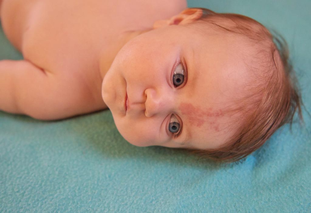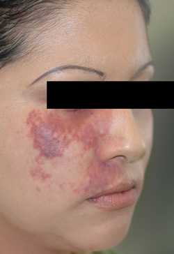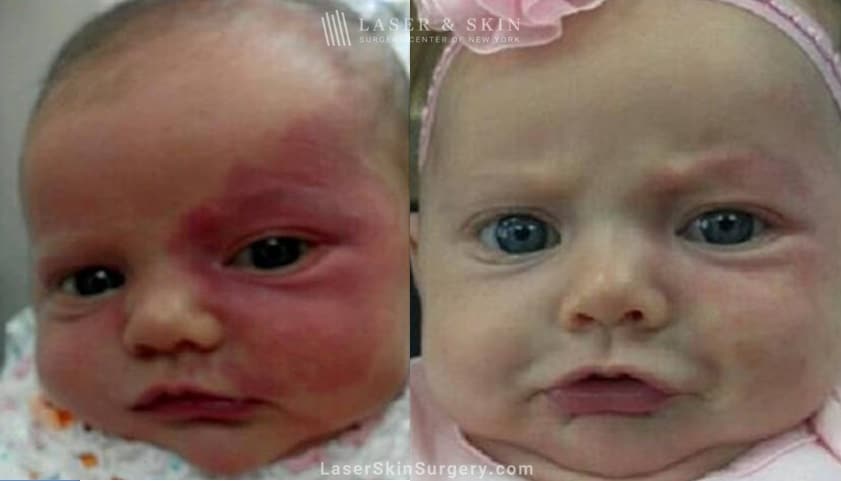


Port-wine stains in the head and neck may develop extracutaneous manifestations causing severe problems. Patients with gingival affection and recurrent otitis were treated by local ear care. Recurrent epistaxis and nasal obstruction due to affected inferior turbinate were treated by Nd:YAG laser therapy, and globus sensation and dysphonia by speech therapy. Cases with lower lip hypertrophy were treated by conventional surgical approaches. Major clinical features included macrocheilia in three cases, gingival bleeding in two, dysphonia with globus sensation, painful parotideal swelling with recurrent otitis, painful lingual swelling, recurrent epistaxis, and nasal obstruction in one case each. Nine patients, mean age 50.4 years, with port-wine stains and clinical symptoms due to extracutaneous involvement, were admitted and treated from 2006 to 2009. Records were reviewed for demographic parameters, extent of the lesion, clinical complications, diagnostic measures, previous treatments, ultimate therapeutic approach, and outcome. Figure 1 An untreated facial capillary malformation (port-wine stain) in a 60-year-old man who presented with a complaint of progressive darkening and development of nodularity in his adult years. This study focuses on other extracutaneous anomalies in patients with nevi flammei of the head and neck, giving rise to functional complications.Ī retrospective study was performed on patients with port-wine stains involving the head and neck area. For the treatment of long-standing port-wine stain that is resistant to laser therapy, surgical methods will bring more satisfactory outcomes than traditional laser therapy.It is well known that port-wine stains of the upper part of the face may herald abnormalities of the brain or eye in the form of Sturge-Weber syndrome. After surgical management, most of the outcomes were satisfactory, without complications or recurrence at long-term follow-up.Ī long-standing nodular port-wine stain can convert to a high-flow malformation with an arterial component, and these lesions are different from early-stage port-wine stains. Victoria blue staining of the nodular lesions showed an intermingling of thick-walled vessels with abundant elastic fibers and thin-walled vessels without elastic fibers, which are findings typical of arteriovenous malformations. The nodules developed in 13 patients, and the mean age for nodule onset was 30 years. After a mean follow-up period of 12 years, the outcomes of surgical management were assessed. All specimens obtained from resection were stained with hematoxylin and eosin and Victoria blue for elastic fibers for histomorphologic analysis.

Port wine stain removal skin#
After removal of the vascular lesions, the affected area was reconstructed with a radial forearm free flap or a skin graft depending on cosmetic considerations. Please call our office today at (417) 889-3332 to schedule a consultation with Dr. Pre-authorization for insurance coverage will be filed if appropriate. The records of 15 patients with long-standing port-wine stain were reviewed for nodules and associated characteristics. The laser will target the vessels under the skin and decrease redness over time. The authors performed total surgical resections of long-standing port-wine stain in the facial region, and attempted to clarify the histomorphologic changes. Although laser therapy for port-wine stain is a safe treatment modality that has been well-established, the long-standing port-wine stain has a tendency to respond less well to laser treatment. A port-wine stain begins with thin macular lesions and eventually becomes hypertrophic and forms nodules.


 0 kommentar(er)
